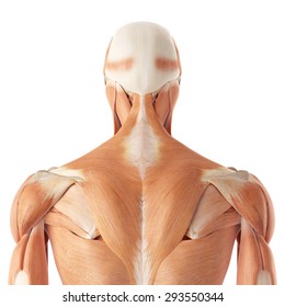
Upper Back Anatomy / Understanding The Anatomy Of The Upper Back Bodyheal. Near the upper surface, toward the back ventral: The iliocostalis muscles are furthest from the spine. All these muscles are therefore associated with movements of the upper limb. The upper back muscles of the rhomboids and the trapezius are responsible for many of the movements of the scapula which in turn plays a huge role in the stability and mobility of the shoulder. The thoracic spine —also referred to as the upper back or middle back—is designed for stability to anchor the rib cage and protect vital internal organs within the chest.
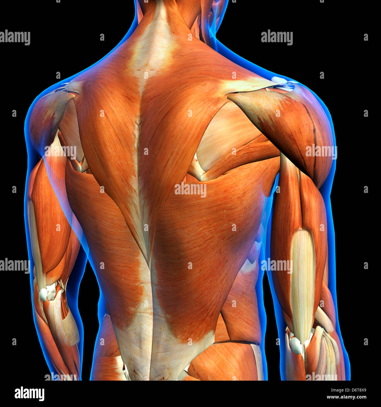
The complexity of this region means that dysfunction can occur either due to injury or progressive pain and degeneration. The nervous system of the thorax is a vital part of the nervous system as a whole, as it includes the spinal cord, peripheral nerves, and autonomic ganglia that communicate with and control many vital organs. The deltoid, teres major, teres minor, infraspinatus, supraspinatus (not shown) and subscapularis muscles (not shown) all extend from the scapula to the humerus and act on the shoulder joint. It connects with the collarbone at the front of the body. There is a set of muscles in the upper back (called the thoracic area) called the spinalis thoracis.
Upper Back Images Stock Photos Vectors Shutterstock from image.shutterstock.com In the upper back, it may cause pain across the group of muscles around the spine, neck, and shoulders. This muscle is located on the upper portion of the back anatomy, underneath the trapezius. This is a tutorial to quickly s. Near, closer to the origin dorsal: The upper back muscles of the rhomboids and the trapezius are responsible for many of the movements of the scapula which in turn plays a huge role in the stability and mobility of the shoulder. In front of, front posterior: The iliocostalis muscles are furthest from the spine. The rib cage also anchors the bones of the head, neck, shoulders, and arms to the trunk of the body.
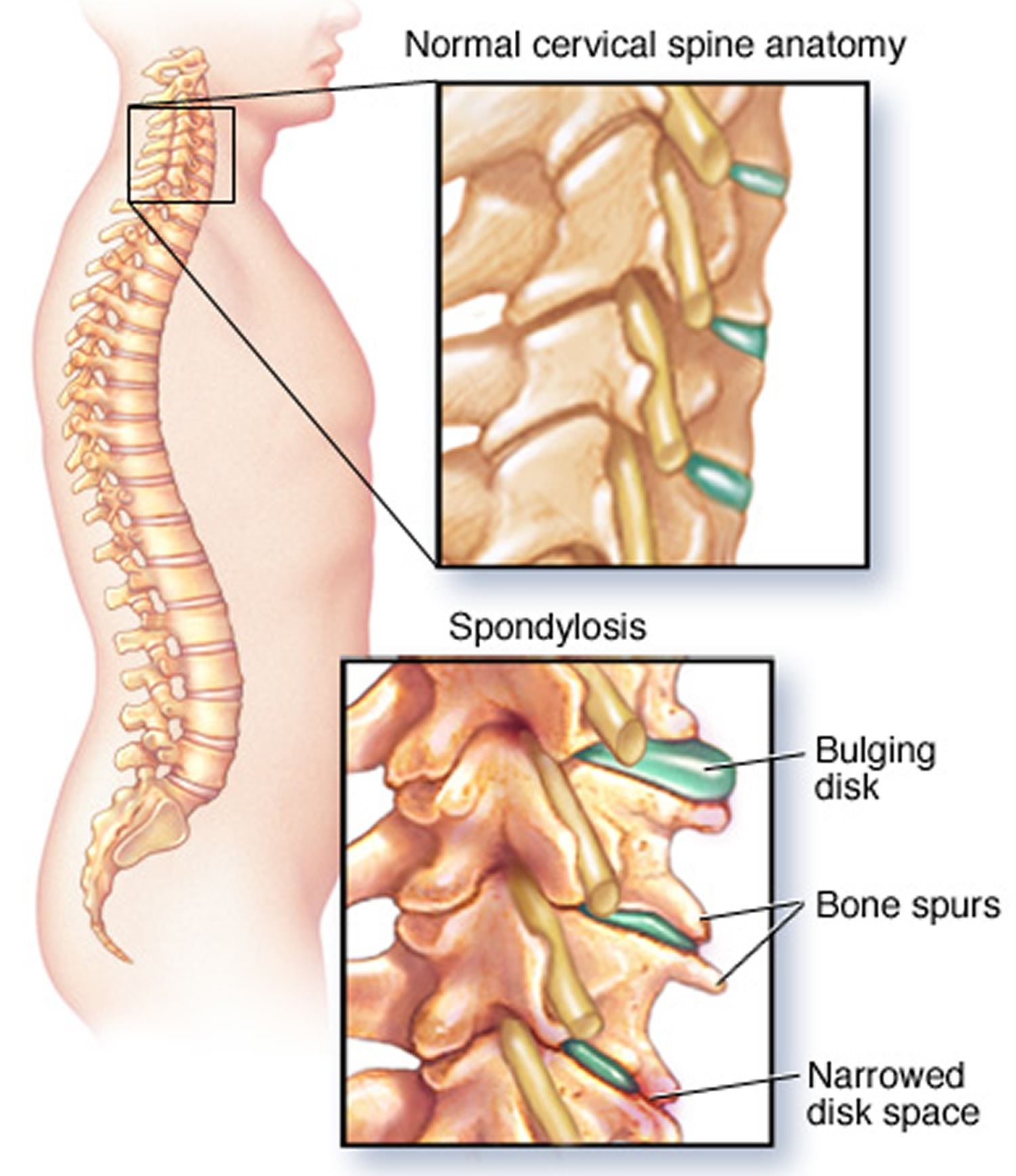
It is like that for several reasons, all of which you can understand by looking at the anatomy of the thoracic spine.
The deltoid, teres major, teres minor, infraspinatus, supraspinatus (not shown) and subscapularis muscles (not shown) all extend from the scapula to the humerus and act on the shoulder joint. Away from, farther from the origin proximal: In front of, front posterior: Related posts of anatomy of the back organs abdominal anatomy kidney. The traps) the latissimus dorsi (a.k.a. The back is the body region between the neck and the gluteal regions. The trapezius and latissimus dorsi muscles connect the upper limb to the vertebral column. It is like that for several reasons, all of which you can understand by looking at the anatomy of the thoracic spine. The rhomboid muscle is activated as you bring and squeeze your scapula or shoulder blades back and together. Some of the exercises which an individual can do to relax or loosen the upper neck and back muscles are: Related posts of upper back muscle diagram muscle relaxation anatomy. It connects with the collarbone at the front of the body. The nervous system of the thorax is a vital part of the nervous system as a whole, as it includes the spinal cord, peripheral nerves, and autonomic ganglia that communicate with and control many vital organs.
It is very stiff, and the thoracic spine has a limited range of motion. Back muscles anatomy here include the trapezius, latissimus dorsi, rhomboid and levator scapulae. They originate from the vertebrae and insert into the scapulae. Muscle relaxation anatomy 12 photos of the muscle relaxation anatomy muscle relaxation anatomy, steps of muscle relaxation anatomy, human muscles, muscle relaxation anatomy, steps of muscle relaxation anatomy These exercises help to loosen up the muscles which facilitate rotation of the spine in either direction to include the erector spinal muscles and the deep spinal muscles.this exercise is especially beneficial for individual who feel stiffness before embarking on sporting.
Upper Back Pain Middle Shoulder Pain Causes Treatment from healthjade.com Study human anatomy with reliable 3d models & detailed articles. Treatment can include laser therapy, steroid injections, lifestyle changes, and massage. Related posts of anatomy of the back organs abdominal anatomy kidney. The deltoid, teres major, teres minor, infraspinatus, supraspinatus (not shown) and subscapularis muscles (not shown) all extend from the scapula to the humerus and act on the shoulder joint. The rhomboid muscle is activated as you bring and squeeze your scapula or shoulder blades back and together. Abdominal anatomy kidney 12 photos of the abdominal anatomy kidney abdomen anatomy kidney, abdominal anatomy kidney, human anatomy, abdomen anatomy kidney, abdominal anatomy kidney. Back muscles anatomy here include the trapezius, latissimus dorsi, rhomboid and levator scapulae. The traps) the latissimus dorsi (a.k.a.
The back functions are many, such as to house and protect the spinal cord, hold the body and head upright, and adjust the movements of the upper and lower limbs.
It comprises the vertebral column (spine) and two compartments of back muscles; In the upper back, it may cause pain across the group of muscles around the spine, neck, and shoulders. Near, closer to the origin dorsal: Rest, medications, physical therapy, and. It runs from the neck to the upper back. It is very stiff, and the thoracic spine has a limited range of motion. The thoracic spine —also referred to as the upper back or middle back—is designed for stability to anchor the rib cage and protect vital internal organs within the chest. The deltoid, teres major, teres minor, infraspinatus, supraspinatus (not shown) and subscapularis muscles (not shown) all extend from the scapula to the humerus and act on the shoulder joint. Related posts of anatomy of the back organs abdominal anatomy kidney. Both the deltoid and the trapezius are firmly attached to the spine of the scapula. Anatomy of muscles in the body The cervical spine supports the weight and movement of your head and protects the nerves exiting your brain. It connects with the collarbone at the front of the body.
Anatomy of back muscles your back consists of three distinct layers of muscles, namely the superficial layer, the intermediate layer, and the deep layer. The complexity of this region means that dysfunction can occur either due to injury or progressive pain and degeneration. The trapezius and latissimus dorsi muscles connect the upper limb to the vertebral column. Abdominal anatomy kidney 12 photos of the abdominal anatomy kidney abdomen anatomy kidney, abdominal anatomy kidney, human anatomy, abdomen anatomy kidney, abdominal anatomy kidney. The iliocostalis muscles are furthest from the spine.
Male Upper Back Muscles Anatomy In Blue X Ray Outline Full Color 3d Computer Generated Illustration On Black Background Stock Photo Alamy from c8.alamy.com Anatomy of muscles in the body The iliocostalis muscles are furthest from the spine. The main superficial muscles of the back are the following: The upper back originates at the base of your neck, incorporates both shoulders and extends down to mid spine, including your ribs. The superficial back muscles are situated underneath the skin and superficial fascia. Anatomy of back muscles your back consists of three distinct layers of muscles, namely the superficial layer, the intermediate layer, and the deep layer. Muscle relaxation anatomy 12 photos of the muscle relaxation anatomy muscle relaxation anatomy, steps of muscle relaxation anatomy, human muscles, muscle relaxation anatomy, steps of muscle relaxation anatomy All about back and spine.
It is like that for several reasons, all of which you can understand by looking at the anatomy of the thoracic spine.
All about back and spine. Powerful muscles that move the head and arms attach to these bones as well. The upper back originates at the base of your neck, incorporates both shoulders and extends down to mid spine, including your ribs. The main superficial muscles of the back are the following: Explore every muscle, bone and organ in 3d It comprises the vertebral column (spine) and two compartments of back muscles; The nervous system of the thorax is a vital part of the nervous system as a whole, as it includes the spinal cord, peripheral nerves, and autonomic ganglia that communicate with and control many vital organs. In the upper back, it may cause pain across the group of muscles around the spine, neck, and shoulders. This is a tutorial to quickly s. It consists of seven vertebrae. All these muscles are therefore associated with movements of the upper limb. Some of the exercises which an individual can do to relax or loosen the upper neck and back muscles are: There is a set of muscles in the upper back (called the thoracic area) called the spinalis thoracis.